Electroencephalography (EEG)
Electroencephalography (EEG) can be used to measure and graphically display the electrical activity of the brain.
What is electroencephalography (EEG)
The term EEG stands for electroencephalography and refers to an examination technique in which the electrical activity of the cerebral cortex is measured. To do this, the neurologist sticks electrodes to the patient’s scalp according to a fixed pattern and connects them together. The measured electrical activity is generated by the discharge of nerve cell clusters.
Electroencephalography (EEG) — procedure
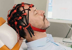
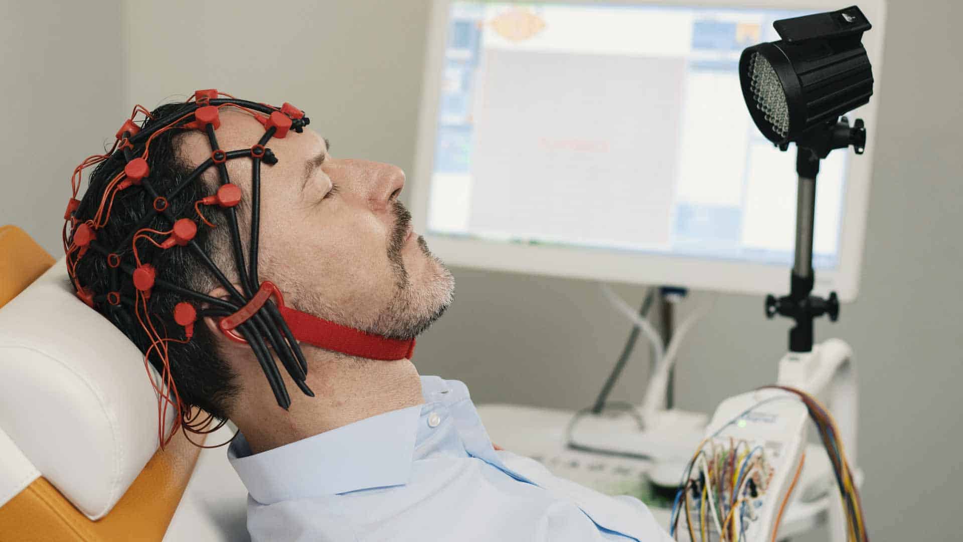
The patient is examined and informed by a neurologist. Up to 21 electrodes are required for a routine EEG, which are usually incorporated into a type of cap. This facilitates placement and adhesion to the patient’s head. The electrodes are coated with a contact gel, attached to the patient’s scalp according to a standardized scheme and connected via cables.
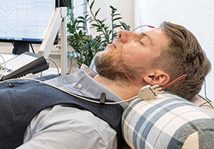
During the actual measurement, which cannot be felt, the patient should be as relaxed and calm as possible and keep their eyes closed. The attending physician gives brief instructions from time to time, for example to open the eyes or solve a simple math problem. This leads to a change in brain activity and is recorded in the EEG. The entire measurement usually takes no longer than 20 to 30 minutes. The electrode cap is then removed.